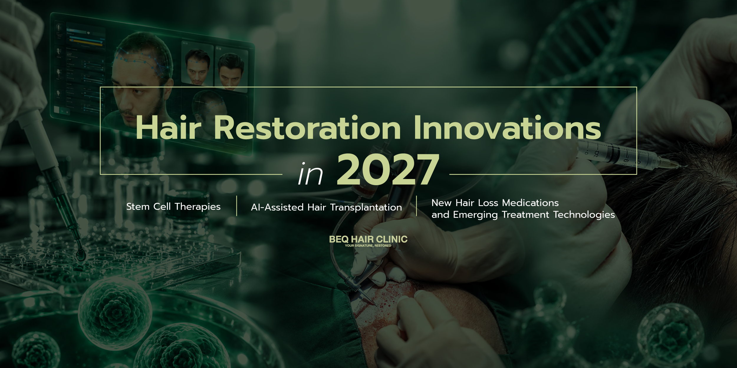
Traditionally, clinical assessment and diagnosis relied predominantly on the physician’s experience, visual judgment, and manual examination. Today, AI has emerged as an important adjunct for hair-restoration physicians around the world. The essence of this transition can be summarized in one statement: “AI not only enables clinicians to visualize hair and scalp structures more clearly; it also converts clinical images into quantitative datasets that can be measured, compared, and followed longitudinally in a scientifically reproducible manner.”
This article explores the integration of advanced technologies—particularly Computer Vision and Deep Learning—with established medical principles to enhance diagnostic precision, treatment planning, and standards of care in hair restoration. To appreciate the significance of AI in this field, it is important first to understand conventional assessment methods and the limitations that hair-restoration physicians have faced for decades prior to the introduction of AI.
Traditional Hair and Scalp Assessment: Methods and Limitations
1. Hair Loss Pattern Assessment
The initial evaluation begins with a detailed medical history and global visual inspection of the scalp to determine the pattern and severity of hair loss. Commonly used classification systems include the Norwood–Hamilton scale for male-pattern hair loss and the Ludwig scale for female-pattern hair loss. These systems are useful for providing an overall assessment of disease severity; however, visual inspection alone cannot reliably characterize subtle alterations in individual hair shafts or microscopic abnormalities that may not be evident to the unaided eye.
2. Physical Tests
Physicians may perform manual tests to assess hair shedding and shaft integrity, including:
- Pull Test: A small bundle of approximately 50–60 hairs is gently grasped and pulled. If more than approximately 10% of the hairs are extracted, the result may be considered abnormal in the appropriate clinical context.
- Tug Test: Approximately 10–20 hairs are held with both hands and gently pulled in opposite directions to assess hair-shaft fragility and breakage.
A limitation of these tests is their inherent variability. Results may be influenced by the examiner’s technique, including the amount of traction applied, which can reduce inter-examiner reproducibility and standardization.
3. Early Dermoscopy / Trichoscopy
In the late 1990s, the dermatoscope—originally developed for the examination of cutaneous lesions—began to be applied to scalp and hair assessment, leading to the development of trichoscopy. Early devices typically provided approximately 10–20× magnification, allowing clinicians to identify findings that may be difficult to appreciate with the naked eye, such as hair-shaft miniaturization, perifollicular erythema, or scaling. In preoperative hair-transplant evaluation, a trichoscope may also be used to examine the donor area and estimate follicular-unit density. Traditionally, follicular units are manually counted within defined areas, such as one square centimeter, and the measurements are averaged to estimate the number of grafts that may be safely available for transplantation.
Nevertheless, hair and scalp assessment in the pre-AI era remained subject to several limitations that could affect diagnosis and treatment planning, including:
- Visual Bias: Estimation of follicular-unit density may depend on subjective clinical judgment. Two hair-transplant physicians may therefore grade disease severity or estimate the number of grafts required differently in the same patient, resulting in inter-rater variability.
- Late Detection: Androgenetic alopecia (AGA) is characterized by progressive hair miniaturization. By the time visible thinning becomes obvious to the naked eye, a substantial proportion of affected hairs may already have undergone advanced miniaturization. Earlier recognition of these changes may provide an opportunity for timely medical intervention.
- Graft-Count Mismatch: Before hair-transplant surgery, physicians must evaluate donor density and estimate the number of follicular units that can be harvested without producing cosmetically significant donor depletion. Manual trichoscopic counting can be time-consuming and visually demanding, particularly in long or overlapping hair. Inaccurate estimation may contribute either to excessive harvesting with visible donor thinning (over-harvesting) or to under-harvesting that limits the ability to achieve an appropriate recipient-site density.
As hair-restoration medicine encountered the practical limitations of human visual assessment, digital technologies increasingly emerged to address these gaps. Since approximately 2015, rapid advances in artificial intelligence—particularly Computer Vision and Deep Learning—have transformed many areas of medical imaging.
A key technological foundation is the Convolutional Neural Network (CNN), a class of deep neural network designed to process grid-like data such as images and video. When trained on large datasets of scalp and hair images, CNN-based systems can perform tasks such as Object Detection, allowing the algorithm to identify and classify relevant visual structures.
Such systems can distinguish among “hair shafts,” “follicular openings,” and the “scalp surface,” even in complex images in which long hairs overlap. When AI is combined with high-resolution digital trichoscopy and polarized illumination to reduce scalp-surface reflection, clinical scalp images can be transformed into quantitative data within seconds, enabling numerical analysis rather than purely visual interpretation.
To illustrate how these technologies may be applied in clinical practice, TrichoLAB provides a useful example. TrichoLAB is an internationally used platform developed through collaboration between FotoFinder Systems, a German company specializing in medical and dermatologic imaging technology, and experts in hair and scalp analysis. In Thailand, this technology is available at a limited number of centers, including BEQ Hair Clinic, where it is offered as FollicleForge AI by TrichoLAB Space Studio. The system demonstrates several ways in which digital analysis can enhance preoperative hair-restoration assessment.
FollicleForge AI by TrichoLAB: Advanced Hair Analysis and 3D Transplant Planning

1. Micron-Level Precision

One of the principal strengths of AI-assisted digital analysis is spatial quantification. Using the dedicated TrichoLAB trichoscopy system, images can be analyzed to estimate the diameter of individual hair shafts in micrometers (µm) and to classify follicular units as single-, double-, or multiple-hair units per square centimeter. Quantitative measurements can assist the physician in identifying hair miniaturization at an earlier stage and may support timely initiation or adjustment of medical therapy when clinically indicated.
2. Safe Donor Area Assessment

In Follicular Unit Excision (FUE), follicular units are harvested from the donor region, typically in the occipital and parietal scalp, where hairs are relatively resistant to androgen-mediated miniaturization. TrichoLAB’s 3D Transplanner software can map the donor region and generate a three-dimensional model incorporating quantitative measurements of donor density and estimated follicular-unit availability. This information can assist the physician in estimating a sustainable harvesting plan and in demonstrating to the patient how different harvesting strategies may affect the remaining donor appearance, thereby helping reduce the risk of unnecessary or excessive extraction (over-harvesting).
3. Same-Spot Tracking
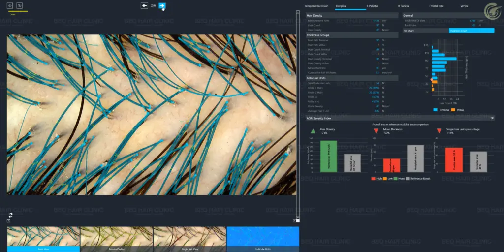
A common challenge in evaluating treatment response—whether after medical therapy or hair transplantation—is ensuring that serial photographs truly represent the same scalp location. Even minor differences in camera angle, illumination, or positioning may create misleading visual impressions. AI-assisted pattern-recognition systems, such as TrichoLAB’s Virtual Tattoo technology, can recognize the patient-specific distribution of follicular openings, effectively creating a follicular map analogous to a biometric pattern. When the patient returns for follow-up at 6 or 12 months, the system can guide image acquisition back to the same scalp location, allowing objective comparison of parameters such as hair count and hair-shaft diameter over time. This improves the reproducibility, transparency, and audit-ability of longitudinal treatment assessment.
4. Non-Invasive Analysis Without Hair Cutting or Shaving

Conventional trichoscopy may be more difficult in patients with longer hair because overlapping shafts can interfere with follicular-unit counting and density calculations. Historically, clipping or shaving may sometimes have been required to obtain more standardized measurements. Current AI-assisted approaches, including TrichoLAB F-Mapping (No-Loss Hair Detection), are designed to account for hairs that are partially obscured in images acquired from different angles. This may enable quantitative assessment of follicular density without requiring routine shaving or plucking, thereby reducing patient anxiety and improving convenience during evaluation.
5. Standardization of Clinical Assessment

Patients with hair loss may consult several hair-transplant physicians and receive different estimates of the number of grafts that can or should be harvested. Such variation may create uncertainty regarding which recommendation is most appropriate. Quantitative analysis using a standardized system such as TrichoLAB can provide a consistent diagnostic baseline for both the physician and the patient, supporting more reproducible calculations and treatment planning when the same methodology and imaging protocol are used.
Despite the increasing ability of AI to “count” and “measure” physical structures, many forms of hair loss still require clinical interpretation by a physician experienced in hair and scalp disorders. Hair loss may result from a broad differential diagnosis, including thyroid disease, alopecia areata, iron-deficiency anemia, syphilis, systemic lupus erythematosus (SLE), severe psychological or physiologic stress, trichotillomania, and many other conditions. AI cannot independently obtain a complete medical history, perform a comprehensive differential diagnosis, interpret laboratory investigations in clinical context, or assess the patient’s psychological status. Therefore, the quantitative data generated by AI must ultimately be interpreted by a qualified clinician in order to determine the underlying cause of the patient’s hair-loss problem.
If you are interested in a hair transplant, or if you have concerns regarding the health of your hair or scalp, please feel free to contact us for further information:
📞 094-441-4965
📩 Line: @beq_haircenter
Click: http://line.me/ti/p/@beq_haircenter
Facebook: BEQ Hair Center
Instagram: beqhaircenter_dr.danai
Instagram: beq_hairclinic
YouTube: BEQ Hair Center
TIKTOK: beq_hair_center